
When a wrist fracture heals out of position
A distal radius malunion is a fracture near the wrist that has healed in an altered position. This can change the relationship between the radius, ulna and wrist joint, contributing to pain, reduced movement or difficulty using the hand. Treatment depends on your symptoms and function, not simply the appearance of an X-ray.
For selected complex injuries, Dr Melsom can arrange patient-specific 3D planning, custom surgical guides and a custom plate. The aim is to restore the bone’s alignment as accurately as practical and improve the conditions for wrist and forearm function. The plan is designed around the individual deformity and the patient’s needs.
Planning the correction in three dimensions
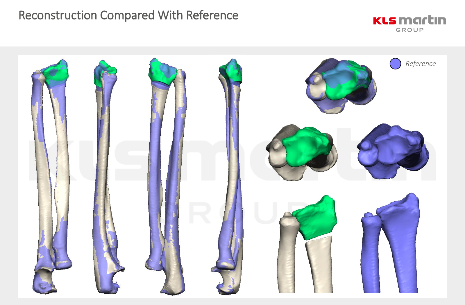
CT scans can be used to build a three-dimensional model of the bones. This lets the surgeon assess shortening, angulation and rotation together, and plan a corrective osteotomy: cutting and repositioning a bone that has healed in the wrong position. Where appropriate, the other forearm can provide a reference for the intended alignment.
Custom guides: transferring the plan to surgery
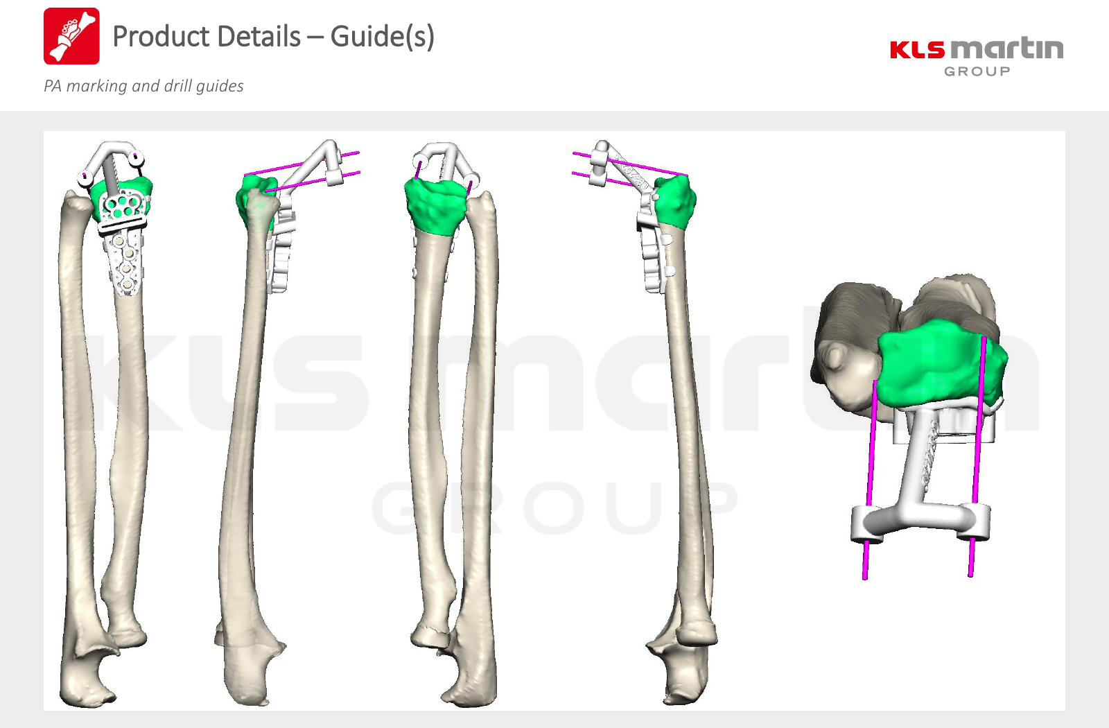
A patient-specific guide is designed to fit the contours of the individual bone. It helps locate planned drill holes and bone cuts, bringing the digital plan into the operation. This can be particularly useful when a deformity involves several directions and is difficult to appreciate on ordinary two-dimensional X-rays. The surgeon still checks the fit, alignment and fixation during surgery.
A plate designed for the planned anatomy
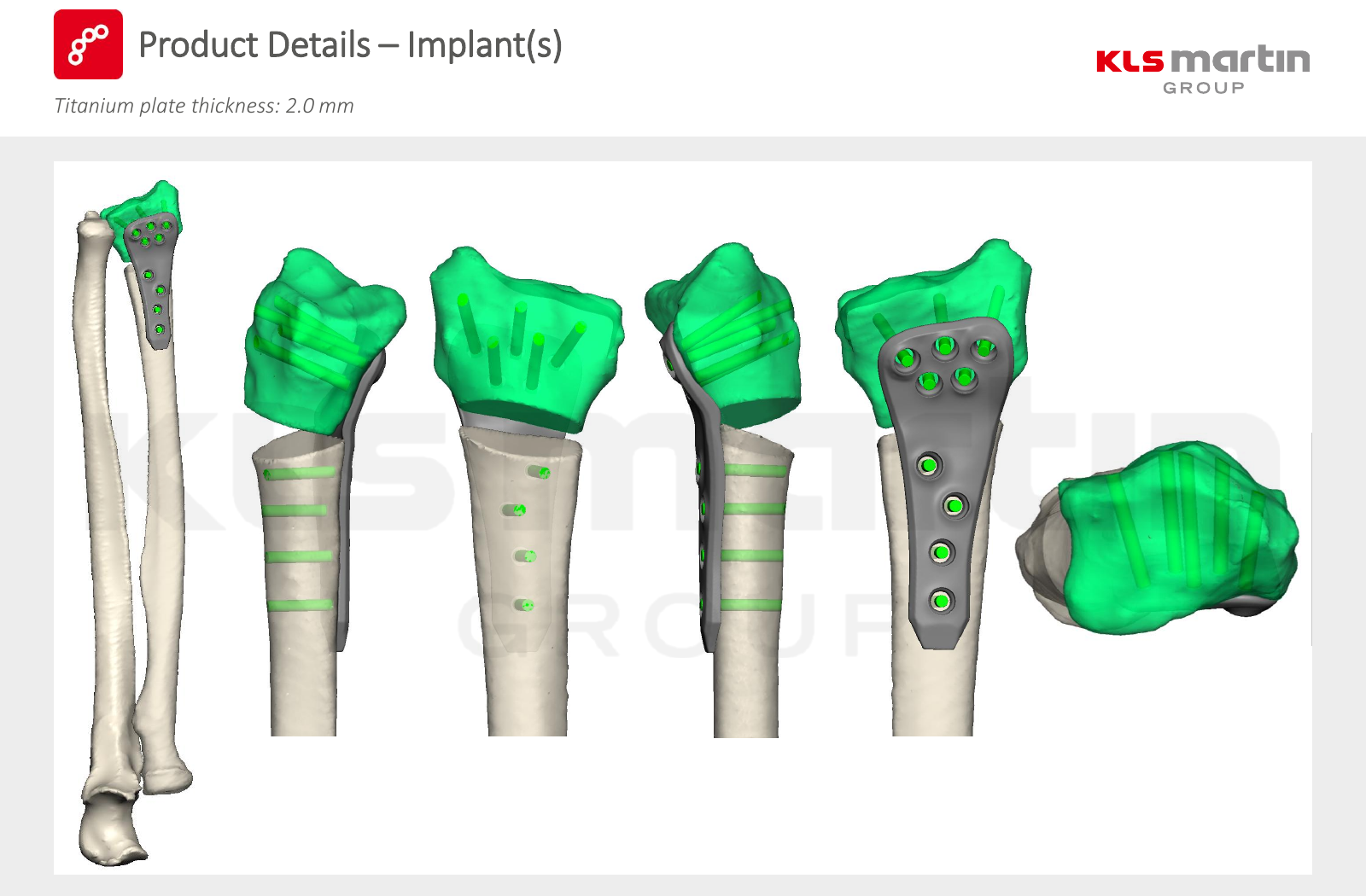
For some reconstructions, a custom titanium plate can be designed to match the intended corrected shape of the bone and the planned screw positions. The plate holds the repositioned bone while it heals. Bone graft may also be needed, depending on the correction. A custom implant is not necessary for every malunion; a standard plate may be appropriate in other cases.
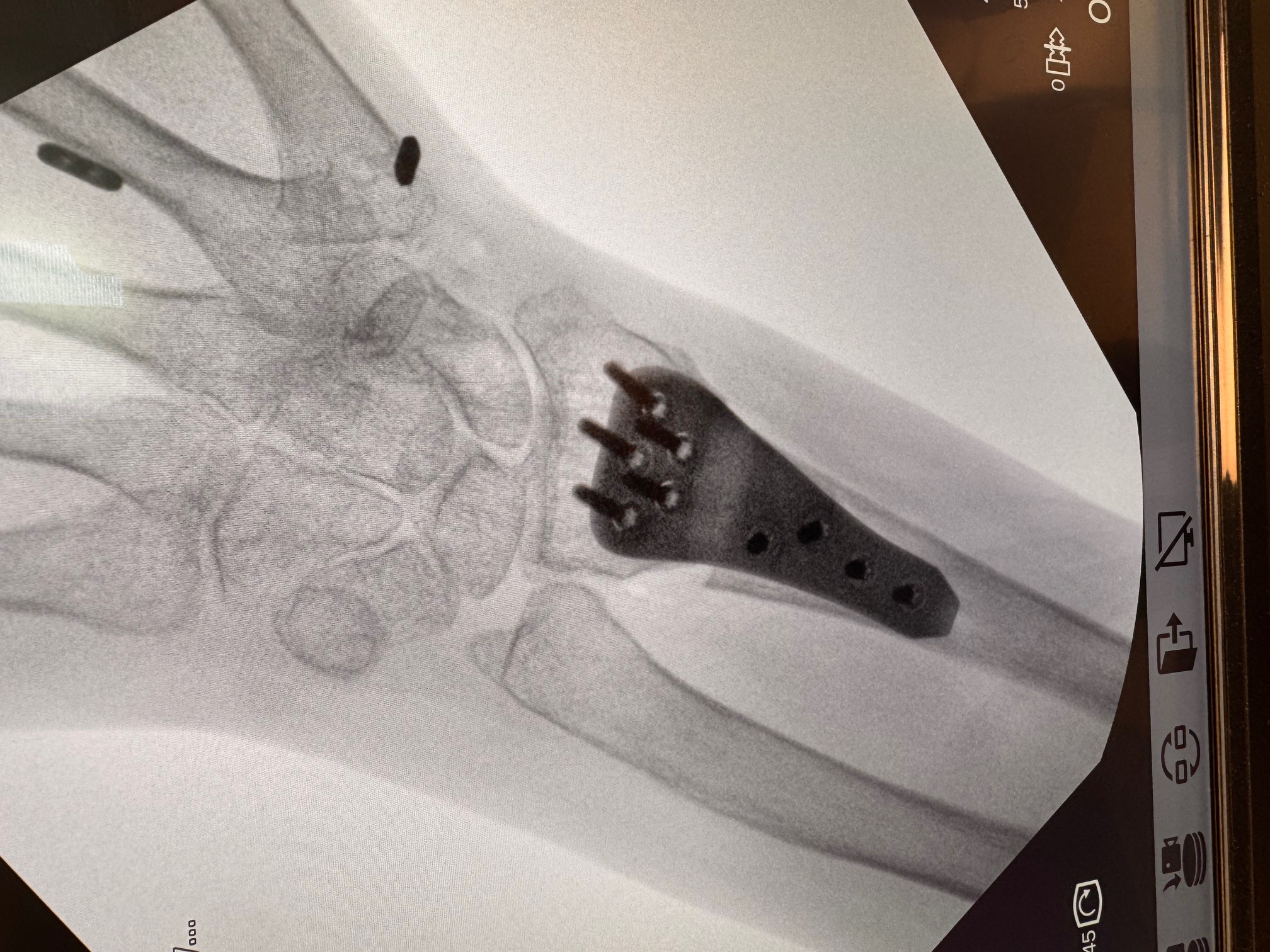
Intra-operative X-rays from the same case
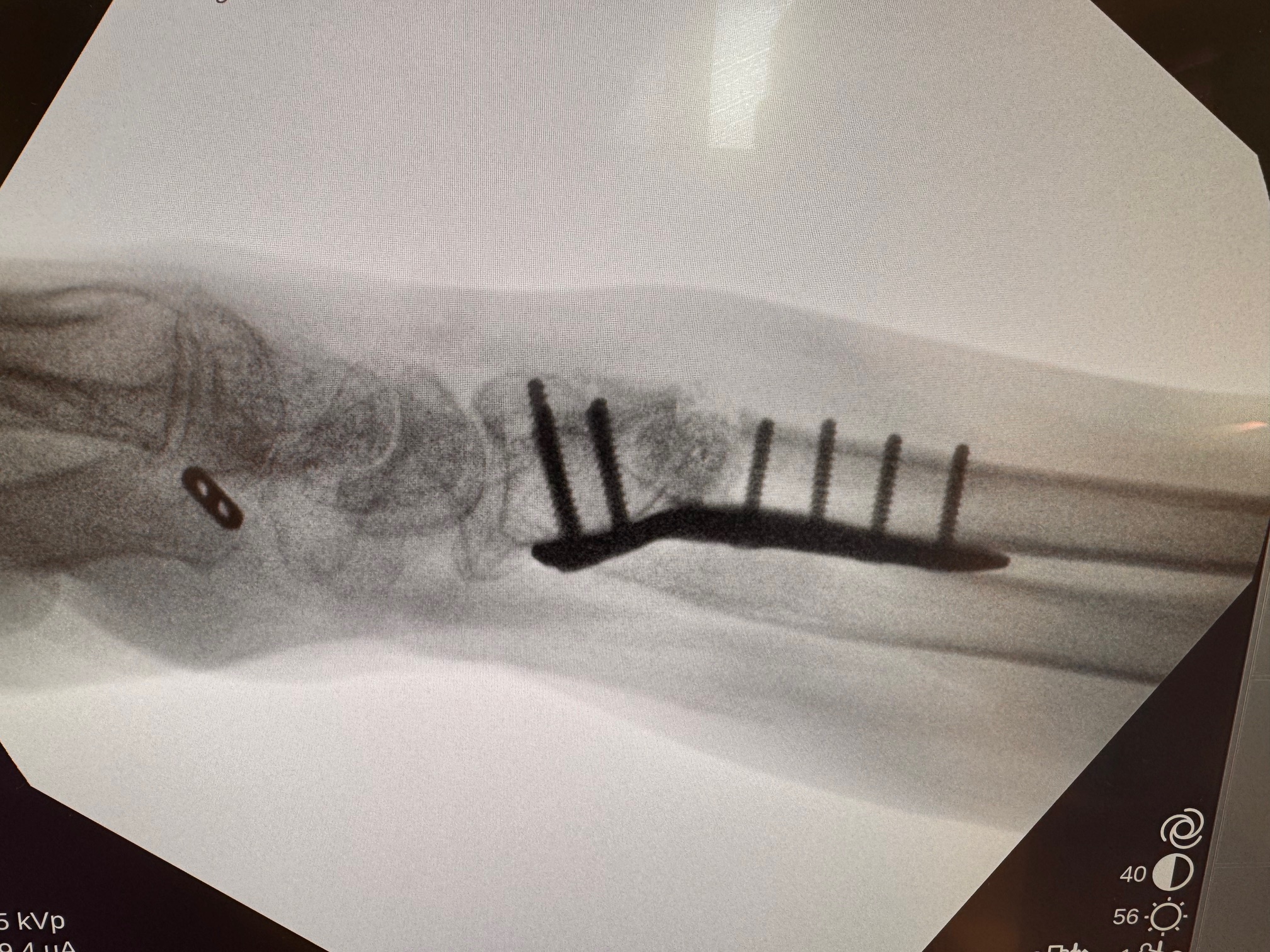
These X-rays were taken during the operation for the case illustrated in the planning images above. They show the plate and screws in position from two different views. The digital images above show the intended reconstruction; these X-rays illustrate its appearance during surgery. They do not show later bone healing or the patient’s eventual movement and function.
What this approach can offer — and its limits
The value is in understanding a complex deformity in three dimensions and preparing guides and fixation for that particular anatomy. Improved alignment can support better mechanics, but restoring alignment does not guarantee normal movement, strength or freedom from pain. Cartilage damage, stiffness, tendon or ligament injury and the ability of the bone to heal also affect recovery.
Published studies describe encouraging results with 3D-planned corrective surgery, but the evidence includes small case series and does not establish that a custom plate is better for every patient. Planning and manufacture add time and cost, and CT imaging involves radiation. Risks include infection, delayed or failed bone healing, nerve or tendon injury, persistent stiffness and irritation from the plate or screws; further surgery may be necessary.
Assessment and rehabilitation
Dr Melsom will review your symptoms, previous treatment, examination and imaging before discussing whether observation, therapy, conventional corrective surgery or a custom reconstruction is appropriate. After surgery, protection and hand therapy are tailored to bone healing and the reconstruction performed. Your return to daily activities and heavier work is guided by follow-up assessment.
Contact the rooms to arrange an assessment or read about hand therapy and rehabilitation.